
Start using our platform today for free and enhance your learning experience with seamless document editing!
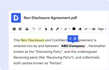
If you often deal with documents kept in your Google Drive, DocHub is a superb option for you to easily and quickly complete, adjust, and approve them. This editor integrates well with Google services, so you can export your which diagrams in model 1 represent the following states for embedded protein b from your Google Drive to the editor without the need of downloading and re-uploading it. Right-click on your document, choose Open With → DocHub PDF Sign and Edit. In our editor, add and assign Signature Fields for all people involved, then click on the Menu button above → Send → select how you want to share your form.
When you edit your neuron function pogil answer key quizlet with DocHub, you merely need a steady web connection and virtually any web browser installed on your device. No need to set up any third-party software or study guides. Simply open a tab with DocHub, drag and drop your file, and edit it immediately.
At DocHub, your data security is our priority. We follow HIPAA, SOC2, GDPR, and other standards, so you can work on your documents with confidence.
Learn more