

Start using our platform today for free to easily fill out and manage your EyeScreen Photographic Examination!
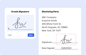
Log in to your DocHub account and add your EyeScreen Photographic Examination - Vision Source Mandan to our editor using one of its upload methods - from your device, cloud storage, protected URL, or your DocHub folders if you have already processed your document before. Open our editor, click the Sign button in the upper toolbar, and choose your signing method. You can upload a picture of your handwritten signature, draw it, type in your name, or utilize a QR code instead.
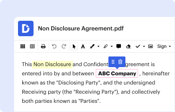
You may easily fill out, edit, and even eSign your EyeScreen Photographic Examination - Vision Source Mandan utilizing DocHub. Create a new account and start your free trial. After that, you may upload the document with the form and easily make all the required edits. No need to print on paper or use a third-party application to sign it, as you can place your electronic signature on your document quicker through DocHub.
At DocHub, your data security is our priority. We follow HIPAA, SOC2, GDPR, and other standards, so you can work on your documents with confidence.
Learn more