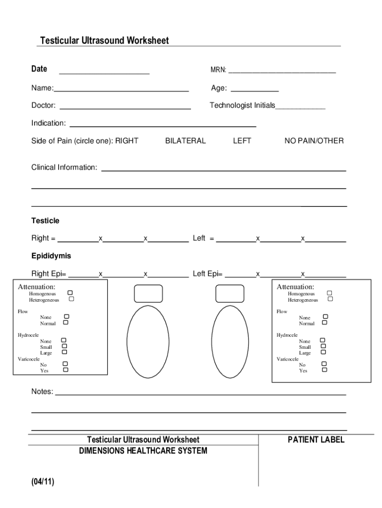


Definition & Meaning
The Testicular Ultrasound Worksheet - Netsolhost is a specialized document used in the medical field to record comprehensive details and findings related to testicular ultrasound examinations. It serves as a structured form that captures vital information about a patient’s condition, clinical observations, and diagnostic results. This worksheet is primarily utilized by healthcare professionals during the assessment of testicular abnormalities, providing a systematic approach to document patient data, ultrasound indications, and findings. The careful documentation aids in ensuring accurate diagnoses and consistent patient care.
Key Elements of the Testicular Ultrasound Worksheet - Netsolhost
The Testicular Ultrasound Worksheet consists of several key components that facilitate the thorough documentation of a testicular ultrasound examination:
-
Patient Details: Includes fields for patient identification information such as name, age, and medical record number. Accurate patient identification ensures that the ultrasound findings are correctly attributed.
-
Clinical Indications: This section outlines the reasons for performing the ultrasound, such as pain, swelling, or suspected abnormalities. Documenting the clinical indications provides context for the findings.
-
Ultrasound Findings: Provides space to record observations regarding the condition of the testicles and surrounding tissues, including the presence of hydrocele, varicocele, or other anomalies. Detailed findings are crucial for diagnosis and treatment planning.
-
Imaging Descriptions: Incorporates fields for describing the characteristics of the ultrasound images, such as echotexture and vascularity. This information helps in differentiating between normal and pathological conditions.
How to Use the Testicular Ultrasound Worksheet - Netsolhost
The worksheet is utilized by following a systematic process to ensure all critical information is captured accurately:
-
Patient Preparation: Before beginning the ultrasound, gather and record the patient's demographic and clinical information on the worksheet to establish a baseline.
-
Clinical Assessment: Use the worksheet to document the initial clinical assessment and indications for the ultrasound examination. This includes noting any symptoms such as pain or swelling.
-
Ultrasound Performance: During the ultrasound, utilize the worksheet to make detailed notes on the findings and observations. This includes the evaluation of both testicular and epididymal structures.
-
Review & Analysis: After completing the ultrasound, review the findings documented on the worksheet and analyze them in the context of the initial clinical indications.
-
Report Generation: Use the documented information from the worksheet to generate a comprehensive ultrasound report for the patient’s medical record.
Steps to Complete the Testicular Ultrasound Worksheet - Netsolhost
Completing the worksheet systematically ensures that no detail is overlooked:
-
Filling Patient Information: Start by entering all necessary patient information, ensuring accuracy to avoid any mix-up with medical records.
-
Documenting Indications for Test: Clearly outline why the ultrasound is being conducted. Include symptoms, medical history, or suspected conditions.
-
Recording Observations: During the examination, systematically note the ultrasound findings such as size, shape, and structure of the testicles, as well as any anomalies.
-
Reviewing Observations: Once the examination is complete, review all entries for completeness and accuracy. Ensure that every relevant detail has been captured.
-
Finalizing the Document: Confirm that all sections are completed and make any necessary corrections before finalizing the worksheet.
Who Typically Uses the Testicular Ultrasound Worksheet - Netsolhost
This worksheet is primarily used by:
-
Radiologists and Sonographers: These medical professionals are responsible for conducting the ultrasound and recording their findings on the worksheet.
-
Urologists: Specialists who may use the worksheet to evaluate the ultrasound results in conjunction with other clinical data to make informed diagnostic and treatment decisions.
-
General Practitioners: Although less specialized, they may utilize the completed worksheet to monitor ongoing conditions affecting the testicles as part of a broader patient care plan.

Important Terms Related to Testicular Ultrasound Worksheet - Netsolhost
Understanding the terminology associated with this worksheet is crucial for its effective use:
-
Hydrocele: A fluid-filled sac around a testicle that results in swelling.
-
Varicocele: An abnormal enlargement of the veins within the scrotum.
-
Echotexture: The texture of tissue as seen on an ultrasound, indicating various conditions based on the appearance.
-
Doppler Ultrasound: A test that uses high-frequency sound waves to measure blood flow.
Examples of Using the Testicular Ultrasound Worksheet - Netsolhost
Several examples illustrate the practical applications of this worksheet:
-
Case Study A: A patient presents with scrotal pain and swelling. The worksheet captures clinical history and an initial physical examination that guides the ultrasound procedure.
-
Case Study B: A male adolescent with a suspected varicocele undergoes an ultrasound, and the findings are documented in detail on the worksheet to confirm diagnosis.
-
Case Study C: Routine examination in a patient with a history of testicular cancer requires repeated documentation of ultrasound findings to monitor for recurrence.
Why Should You Use the Testicular Ultrasound Worksheet - Netsolhost
Utilizing the worksheet offers several benefits:
-
Standardization: Ensures consistent documentation across different practitioners and facilities, assisting in maintaining high standards of care.
-
Comprehensive Record: Serves as a thorough record of the testicular evaluation, aiding in longitudinal care and follow-up examinations.
-
Diagnostic Support: Provides essential data that supports accurate diagnosis and treatment planning, enhancing the overall quality of patient care.