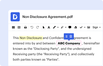
Type text, add images, blackout confidential details, add comments, highlights and more.
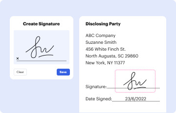
02. Sign it in a few clicks
Draw your signature, type it, upload its image, or use your mobile device as a signature pad.
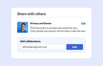
03. Share your form with others
Send information resources via email, link, or fax. You can also download it, export it or print it out.
How to use or fill out Spine Evaluation Form with our platform
Ease of Setup
DocHub User Ratings on G2
Ease of Use
DocHub User Ratings on G2
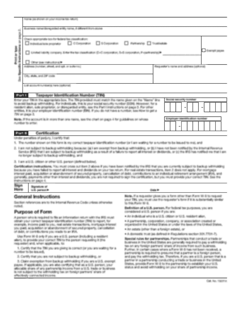
Click ‘Get Form’ to open the Spine Evaluation Form in the editor.
Begin by entering your personal information at the top, including your name, account number, and date. Indicate your dominant hand by checking the appropriate box.
In the 'Chief Complaint' section, specify which part of your spine is concerning you today by selecting from the options provided.
Detail the onset of your symptoms in the designated area. Be as descriptive as possible about when they began and any preceding events.
Use the Pain Diagram to illustrate where you feel pain on your body. Utilize the symbols provided to indicate different types of sensations.
Rate your pain severity over the last two weeks using a scale from 0 to 10 for both back/neck and leg/arm pain.
Complete sections regarding previous treatments, consultations, and any medications tried. This will help provide a comprehensive view of your condition.
Finally, review all entries for accuracy before saving or sharing your completed form directly through our platform.
Start filling out your Spine Evaluation Form online for free today!
The word Cedocore is also coded on the back or spine of the Survivalism physical CD packaging in the form of a sequence of dashes in numbers coresponding toRead more
To request a spine evaluation, please answer the questions below and click on the button that says Submit. We will contact you within one business day.Read more
Cookie consent notice
This site uses cookies to enhance site navigation and personalize your experience.
By using this site you agree to our use of cookies as described in our Privacy Notice.
You can modify your selections by visiting our Cookie and Advertising Notice.