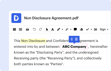
Type text, add images, blackout confidential details, add comments, highlights and more.
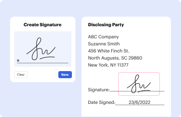
02. Sign it in a few clicks
Draw your signature, type it, upload its image, or use your mobile device as a signature pad.
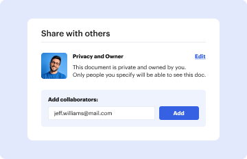
03. Share your form with others
Send ultrasound order form via email, link, or fax. You can also download it, export it or print it out.
How to use or fill out ultrasound order form with our platform
Ease of Setup
DocHub User Ratings on G2
Ease of Use
DocHub User Ratings on G2
Click ‘Get Form’ to open the ultrasound order form in the editor.
Begin by entering the 'Order Date' and the 'Ordering Physician' details at the top of the form. This information is crucial for processing your request.
Fill in the 'Patient Name', 'D.O.B.', and 'Appt Date' fields to ensure accurate patient identification and scheduling.
In the 'Diagnosis/Symptoms' section, provide relevant medical information that justifies the ultrasound procedure.
Select the appropriate study description from the list provided, such as 'US Abdominal Aorta' or 'US Pelvic Sonogram'. Each option corresponds to specific CPT codes listed next to them.
Complete any additional fields like 'Precert Number' and 'Appt Time' as required for your appointment.
Finally, ensure that you have a designated area for the physician's signature before submitting the form.
Start using our platform today to streamline your ultrasound order process for free!
An Ultrasound can be used to see blood flow as well as determine the source of any pain, swelling or infection. This test requires an order from your doctor, nurse practitioner, or physician assistant.
Can you just ask for an ultrasound?
Even though prenatal ultrasounds are safe, you should only have them when its medically necessary. If theres no reason for an ultrasound (for example, if you just want to see your baby), your insurance company might not pay for it.
What is an ultrasound order?
An ultrasound order, is essentially a prescription or referral from a healthcare provider requesting an ultrasound examination for a patient. This order outlines the medical necessity for the ultrasound procedure and provides pertinent information for the radiologist or sonographer performing the scan.
Can I do an ultrasound without a referral?
Importantly, private clinics provide flexible scheduling, same-day appointments, direct patient access and no GP referral required. Whether youre experiencing unexplained symptoms or want to monitor a known condition, a private ultrasound without referral means you get timely, accurate insight without the wait.
Can you refer yourself for an ultrasound?
Whilst you dont need a GP referral to self-refer for a scan, we do require your NHS GP practice details - we will send all reports to your NHS GP in line with good medical practice. Just fill in this quick and easy form, and well be in touch to arrange your scan. Which ultrasound service(s) do you require?
vitco properties
Ultrasound ordering cheat sheetUltrasound request for pregnancyAsante lab order formAsante lab orders
Security and compliance
At DocHub, your data security is our priority. We follow HIPAA, SOC2, GDPR, and other standards, so you can work on your documents with confidence.
You do not need a referral for these scans. Echo Amsterdam is open 7 days and 3 evenings a week. That way you can plan your appointment at a day and time that fits your, and your partners, schedule and in the case of a (medical) emergency we can fit you in as soon as possible. Here you find our privacy statement.
Do I need a script for an ultrasound?
Yes. A prescription from your physician is always required.
How do I schedule an ultrasound for pregnancy?
Booking a Pregnancy Ultrasound (Sonogram) is easy using LabFinder. Just choose your location and enter your insurance information to find the closest Pregnancy Ultrasound (Sonogram) near you.
Related links
ULTRASOUND REFERRAL ORDER FORM
ULTRASOUND REFERRAL ORDER FORM. FOR NON-EPIC USERS. Please fax required documents prior to scheduling to: 314-747-1637. □ This form □ Insurance card (front andRead more
(Radiology/Nuclear Medicine/Ultrasound/Computed Tomography Examinations) STANDARD FORM 519-B (Rev. 8-83). Prescribed by GSA/MIR FIRMR. (41 CFR) 201Read more
Cookie consent notice
This site uses cookies to enhance site navigation and personalize your experience.
By using this site you agree to our use of cookies as described in our Privacy Notice.
You can modify your selections by visiting our Cookie and Advertising Notice.