


Key Components of a Microscope Fill in the Blank Worksheet
A microscope fill in the blank worksheet is an effective educational tool used to assess and reinforce students' understanding of microscope parts and their functions. This type of worksheet typically includes a diagram of a microscope alongside prompts that require students to label various components.
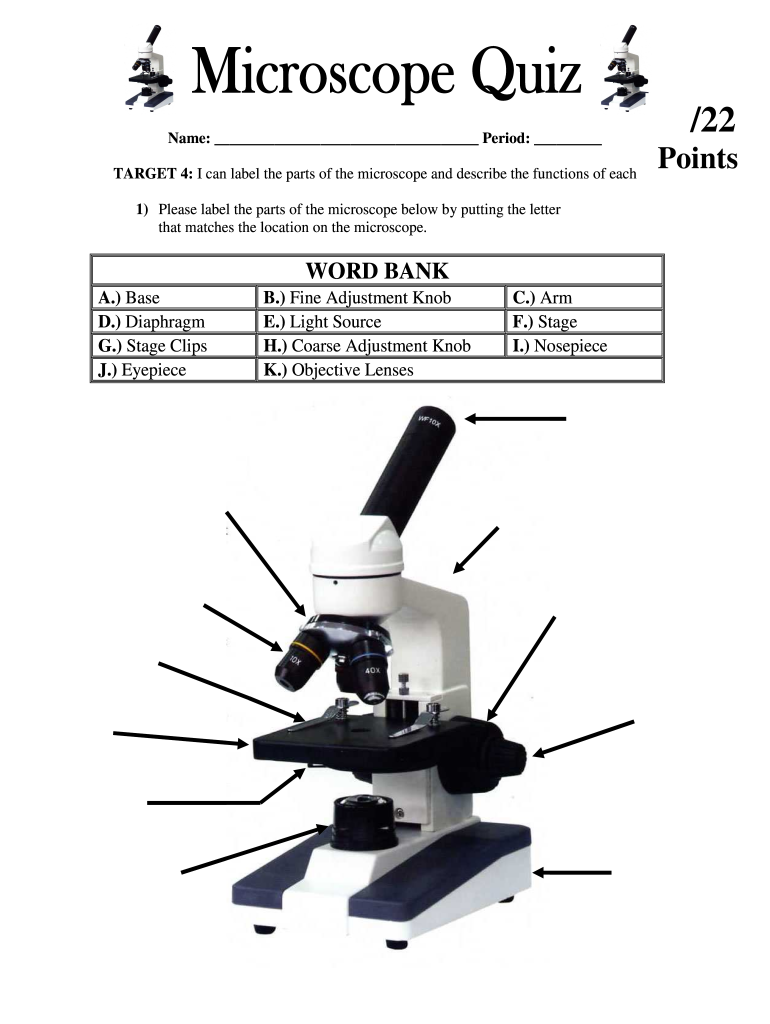
Understanding Microscope Parts
The standard microscope is made up of several key components, each serving a specific function in the microscopy process. Some of the primary parts that may be included in a fill in the blank exercise are:
-
Eyepiece (Ocular Lens): The lens at the top through which the viewer looks. It usually has a magnification of 10x.
-
Objective Lenses: Located on a rotating nosepiece, these lenses range from low (4x) to high (100x) magnification, helping to focus on details of the specimen.
-
Stage: The flat platform where slides are placed for observation. It often has clips to hold the slides in place.
-
Illuminator: Provides light to illuminate the specimen, often adjusted for brightness.
-
Iris Diaphragm: Controls the amount of light reaching the specimen, enhancing contrast and visibility.
Understanding these parts and their functions is essential for students studying biology or related disciplines.
Functions of Microscope Components
Each part of the microscope plays a critical role in the functionality of the device. Knowing these functions helps students complete their fill in the blank activities accurately. Here are several important components and their specific roles:
-
Purpose of the Eyepiece: Magnifies the image received from the objective lens for the viewer.
-
Function of Objective Lenses: Allows for various levels of detail depending on the magnification, facilitating thorough examination of specimens.
-
Role of the Stage: Provides stability and support for slides, enabling precise focus during observations.
-
Importance of the Illuminator: Ensures proper visibility of the specimen, which is crucial for effective microscopy.
-
Significance of the Iris Diaphragm: Adjusts lighting which can impact the clarity and detail of the image seen through the eyepiece.
Educators can structure their worksheets around these concepts to enhance comprehension and retention.
Designing the Fill in the Blank Exercise
Creating a fill in the blank activity involves structuring the worksheet in a way that promotes active engagement. Important elements to include are:
-
Diagram Inclusions: A clear, unlabeled diagram of a microscope enables students to identify and label parts accurately.
-
Word Bank: A list of terms such as "eyepiece," "objective lens," and "stage" can aid students while preventing unnecessary frustration.
-
Task Requirements: Instructions should clarify that students must match terms from the word bank to each component shown in the diagram.
-
Assessment of Knowledge: Include sections that ask for descriptions of each microscope part’s function in addition to labeling to assess both recognition and understanding.
A well-structured fill in the blank worksheet reinforces learning and enables easier recognition of the parts of the microscope in real-world applications.
Benefits of Using Worksheets in Education
Integrating microscope fill in the blank exercises in classrooms provides several educational advantages:
-
Active Learning: Engages students directly with the material, promoting better understanding through hands-on activities.
-
Assessment Tool: Serves as a reliable method for educators to gauge student comprehension of microscope anatomy and functionality.
-
Visual Learning: The use of diagrams caters to visual learners, enhancing retention of complex information.
-
Collaboration: Encourages students to work in pairs or groups, fostering teamwork while discussing and discovering the subject matter together.
These benefits highlight the effectiveness of interactive worksheets in improving educational outcomes, especially in scientific fields.
Examples of Worksheet Implementations
Educators can adopt various approaches when using a fill in the blank worksheet on microscope parts, such as:
-
Individual Assignments: Distributing worksheets for independent completion to assess individual understanding.
-
Group Activities: Organizing students into small groups to facilitate collaborative discussions about microscope functions, followed by filling in the blanks to promote peer learning.
-
Quizzes: Incorporating the worksheet as part of a quiz format to assess knowledge retention after a lesson on microscopes.
-
Thematic Units: Integrating the worksheet into larger units on cell biology or microbiology, reinforcing relevant concepts alongside practical skills.
By implementing these strategies, students can develop a well-rounded understanding of microscopes and enhance their learning experience.
Digital Options for Worksheet Distribution
With the increasing prevalence of technology in education, teachers can also utilize digital formats for microscope fill in the blank worksheets. Options include:
-
PDF Downloads: Allowing easy access for students to print and complete at home or in class.
-
Interactive Online Tools: Websites and applications that enable students to fill in blanks digitally, making the learning process more engaging.
-
Integration with Learning Management Systems: Distributing worksheets through platforms like Google Classroom or Canvas can streamline assignment submissions and grading processes.
These digital tools offer flexibility and accessibility, helping to accommodate diverse learning environments and student needs.
