

Not all formats, including OMM, are designed to be easily edited. Even though a lot of tools can help us tweak all file formats, no one has yet invented an actual all-size-fits-all tool.
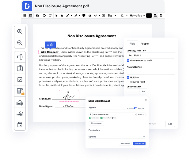
DocHub offers a easy and streamlined tool for editing, managing, and storing paperwork in the most popular formats. You don't have to be a tech-knowledgeable person to work in cross in OMM or make other changes. DocHub is robust enough to make the process easy for everyone.
Our feature allows you to change and edit paperwork, send data back and forth, generate dynamic documents for data gathering, encrypt and protect forms, and set up eSignature workflows. In addition, you can also generate templates from paperwork you utilize frequently.
You’ll find a great deal of additional tools inside DocHub, such as integrations that let you link your OMM file to a variety productivity apps.
DocHub is an intuitive, cost-effective way to handle paperwork and simplify workflows. It provides a wide range of tools, from creation to editing, eSignature solutions, and web document developing. The program can export your files in multiple formats while maintaining greatest protection and adhering to the highest data security standards.
Give DocHub a go and see just how easy your editing process can be.
Muscle contraction is at the basis of all skeletal movements. Skeletal muscles are composed of muscles fibers which in turn are made of repetitive functional units called sarcomeres. Each sarcomere contains many parallel, overlapping thin (actin) and thick (myosin) filaments. The muscle contracts when these filaments slide past each other, resulting in a shortening of the sarcomere and thus the muscle. This is known as the sliding filament theory. Cross-bridge cycling forms the molecular basis for this sliding movement. - Muscle contraction is initiated when muscle fibers are stimulated by a nerve impulse and calcium ions are released. - The troponin units on the actin myofilaments are bound by calcium ions. The binding displaces tropomyosin along the myofilaments, which in turn exposes the myosin binding sites. - At this stage, the head of each myosin unit is bound to an ADP and a phosphate molecule remaining from the previous muscular contraction. - The myosin heads release these pho
