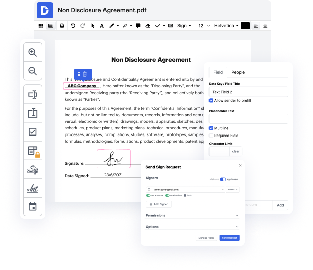


Not all formats, such as ACL, are created to be easily edited. Even though a lot of features will let us tweak all document formats, no one has yet invented an actual all-size-fits-all tool.
DocHub offers a easy and efficient tool for editing, handling, and storing documents in the most widely used formats. You don't have to be a tech-knowledgeable person to wipe out attachment in ACL or make other tweaks. DocHub is powerful enough to make the process simple for everyone.
Our tool allows you to alter and tweak documents, send data back and forth, create dynamic forms for data collection, encrypt and shield paperwork, and set up eSignature workflows. Additionally, you can also create templates from documents you utilize frequently.
You’ll locate a great deal of additional tools inside DocHub, such as integrations that allow you to link your ACL document to a variety productivity applications.
DocHub is a straightforward, cost-effective option to handle documents and streamline workflows. It provides a wide selection of tools, from generation to editing, eSignature professional services, and web form building. The application can export your files in many formats while maintaining greatest safety and following the highest data security criteria.
Give DocHub a go and see just how simple your editing transaction can be.
so this is a somewhat interesting case if we were to look at just the anterior cruciate ligament on this image here we can see the anterior cruciate ligament it seems to go up it doesnamp;#39;t seem to be connecting at the familiar attachment but we may not be entirely sure if we looked at the quality of the ligament itself it may show that it is a little bit thickened and if one didnamp;#39;t have any other information or history then one could think of this even possibly being a mucoid degenerate ACL if we take a quick look at this we can see the TR and TT te times on this R 16 and Iamp;#39;m sorry te and TR times are 16 and 1700 making this although itamp;#39;s a PD weighted image itamp;#39;s um or t1-weighted proton density image if we look at the dedicated proton density image on this you can see the ligament architecture much better but you also see the discontinuity at the femoral attachment but again since you see a vertical ligament one may occasionally be inclined to bel
