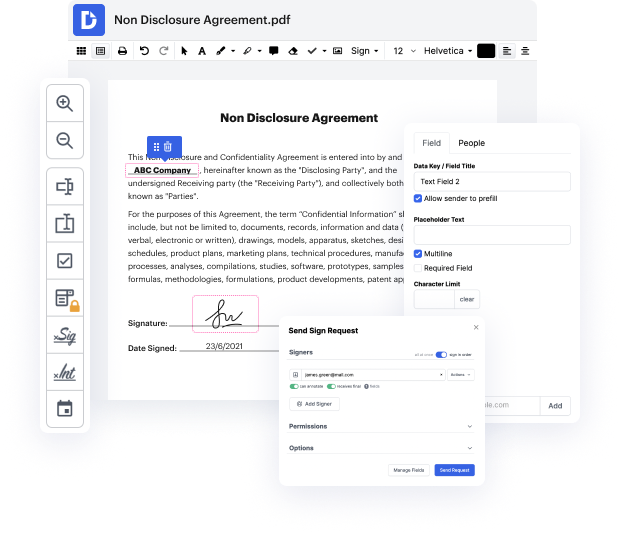


DocHub is an all-in-one PDF editor that enables you to snip chapter in ACL, and much more. You can highlight, blackout, or remove paperwork fragments, add text and pictures where you need them, and collect information and signatures. And since it runs on any web browser, you won’t need to update your software to access its professional capabilities, saving you money. With DocHub, a web browser is all you need to handle your ACL.
Sign in to our service and adhere to these steps:
It couldn't be easier! Improve your document processing now with DocHub!
hello everyone this is Dr Sam and today you will learn about ACL TI on MRI anterior cruciate ligament or ACL is an important liament which helps in stabilizing the knee joint MRI images especially in sagittal planes are very helpful in visualizing the CL we will compare the normal image the normal MRI image of ACL with ACL tiar these are T1 weighted images in sagittal plane this is the knee this bone is the femur and the bone over here is the tibia this bone is the patella we can also see the hofa fat pad appearing hyper intense in D1 images and this is the ACL this fibrous hypointense dark band is the ACL it inserts on the medial aspect of the lateral femoral cile its tibial insertion is on the anterior part of the inter condar Eminence of the tibia at the tibial Plateau so this is the appearance of the ACL we can also see part of the posterior cruciate ligament the PCL in this image the PCL is posterior to the ACL the image on the right shows a complete ACL tier in this case we do no
