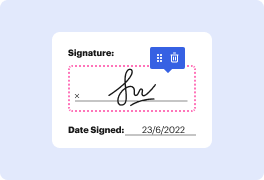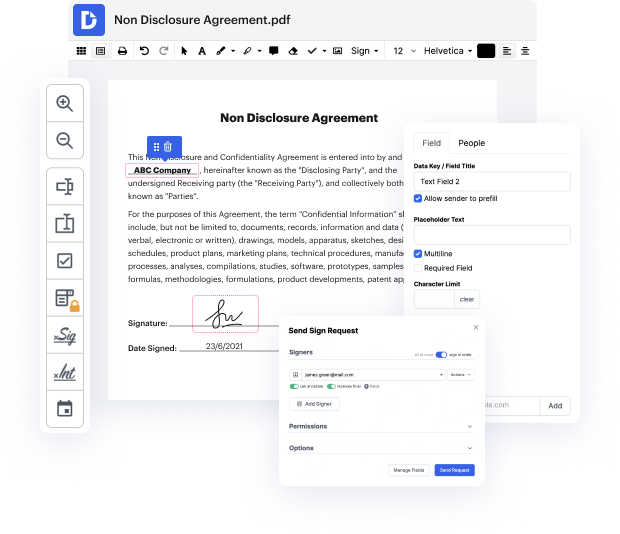


ASC may not always be the easiest with which to work. Even though many editing capabilities are available on the market, not all give a simple tool. We developed DocHub to make editing easy, no matter the document format. With DocHub, you can quickly and effortlessly rework marking in ASC. On top of that, DocHub offers a range of other functionality including form creation, automation and management, sector-compliant eSignature solutions, and integrations.
DocHub also helps you save time by producing form templates from documents that you utilize regularly. On top of that, you can benefit from our numerous integrations that allow you to connect our editor to your most used applications easily. Such a tool makes it quick and easy to deal with your documents without any delays.
DocHub is a handy feature for personal and corporate use. Not only does it give a all-encompassing collection of features for form creation and editing, and eSignature integration, but it also has a range of capabilities that come in handy for creating multi-level and simple workflows. Anything added to our editor is stored risk-free in accordance with leading industry standards that protect users' data.
Make DocHub your go-to choice and simplify your form-driven workflows easily!
this video demonstrates endovascular repair of ascending iotic secular aneurysm in a 78 year old female who presented with a 4.1 centimeter ascending iotic aneurysm thought to be secondary to penetrating iot calcium she complained of chest pain on arrival and has an active diagnosis of pancreatic cancer she was deemed a poor open surgical candidate ct angiogram of the chest demonstrated a 4.1 centimeter ascending aortic secular aneurysm this was secondary to a penetrating iotic ulcer demonstrated here is this ascending iotic circular aneurysm we begin our procedure by gaining access to the right internal jugular vein using a mini stick needle wire and micropuncture sheath we then upsize to a seven french sheath and introduce a temporary pacemaker wire in the right ventricle and this was connected for rapid ventricular placing next the left common femoral artery was excess under ultrasound guidance using a mini stick needle followed by the wire and micropuncture sheath he then upsized t
