
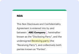


Document generation and approval certainly are a key priority of every organization. Whether handling sizeable bulks of files or a particular contract, you need to remain at the top of your productiveness. Choosing a excellent online platform that tackles your most frequentl papers creation and approval problems might result in quite a lot of work. Numerous online platforms offer merely a minimal set of editing and signature functions, some of which could possibly be useful to manage image formatting. A solution that handles any formatting and task might be a exceptional option when deciding on application.
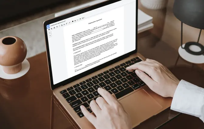
Get file administration and creation to a different level of efficiency and excellence without opting for an difficult interface or costly subscription options. DocHub gives you tools and features to deal effectively with all file types, including image, and carry out tasks of any complexity. Modify, arrange, and produce reusable fillable forms without effort. Get complete freedom and flexibility to correct dent in image anytime and safely store all your complete documents in your user profile or one of many possible integrated cloud storage platforms.
DocHub provides loss-free editing, signature collection, and image administration on the professional level. You do not have to go through exhausting guides and invest a lot of time finding out the platform. Make top-tier secure file editing a regular practice for the every day workflows.
today we are going to demonstrate how a cone cut image is created and how to correct it this will include how to correctly and incorrectly take maxillary anterior periapical image and a bitewing image when taking an anterior maxillary central periapical or pa place the sensor parallel to the central axis of the maxillary incisors and ask the patient to bite down firmly on the bite block then place the pid to the center of the aiming ring to ensure that it is parallel to the indicator arm this is called paralleling technique paralleling technique means the central axis of the root of the tooth is parallel to the placed sensor if paralleling technique cannot be used due to a narrow palate or other interference then you would use a technique called bisecting angle bisecting angle technique is when the central ray is directly perpendicular to the imaginary plane that bisects the angle between the sensor and the root of the tooth this is a great pa because you can see the open mesial conta
At DocHub, your data security is our priority. We follow HIPAA, SOC2, GDPR, and other standards, so you can work on your documents with confidence.
Learn more