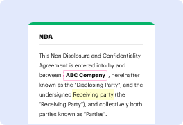


PAP may not always be the simplest with which to work. Even though many editing capabilities are out there, not all give a straightforward solution. We developed DocHub to make editing easy, no matter the form format. With DocHub, you can quickly and easily erase outline in PAP. In addition to that, DocHub provides an array of other functionality such as form creation, automation and management, sector-compliant eSignature solutions, and integrations.
DocHub also lets you save time by producing form templates from paperwork that you use regularly. In addition to that, you can benefit from our a lot of integrations that allow you to connect our editor to your most utilized apps with ease. Such a solution makes it quick and easy to deal with your documents without any slowdowns.
DocHub is a handy feature for personal and corporate use. Not only does it give a all-encompassing suite of capabilities for form creation and editing, and eSignature integration, but it also has an array of capabilities that prove useful for producing complex and straightforward workflows. Anything added to our editor is saved safe according to leading industry criteria that shield users' data.
Make DocHub your go-to option and simplify your form-based workflows with ease!
hi welcome to a lecture about cervical glandular lesions my name is natalie binet today i will discuss the following this is the outline so weamp;#39;ll start with normal under cervix then move on to benign glandular lesions of the cervix and then adenocarcinoma of the uterine cervix so weamp;#39;ll start first with normal and reactive endocervical cells so normal endocervical glands are noted here in an endocervical curettage specimen which honestly is where practicing pathologists encounter these cells very frequently you can show base you can see basally oriented nuclei with a columnar shape and apically oriented mucin reactive forms can also be noted in the endocervical curettage specimens including nuclear enlargement and focal multinucleation which you can see in the center of the photo there drying artifact is also often present and must be interpreted within the context of other cells within the specimen these are important changes to note on histologic section as they can al
