
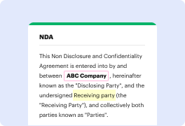


Not all formats, including ACL, are developed to be effortlessly edited. Even though numerous tools can help us edit all file formats, no one has yet created an actual all-size-fits-all solution.
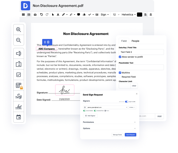
DocHub provides a simple and efficient solution for editing, taking care of, and storing papers in the most popular formats. You don't have to be a tech-knowledgeable person to blot trait in ACL or make other changes. DocHub is powerful enough to make the process straightforward for everyone.
Our feature enables you to change and edit papers, send data back and forth, generate dynamic forms for information collection, encrypt and safeguard forms, and set up eSignature workflows. In addition, you can also generate templates from papers you use frequently.
You’ll locate a great deal of other functionality inside DocHub, including integrations that allow you to link your ACL file to different business apps.
DocHub is a simple, cost-effective option to manage papers and simplify workflows. It offers a wide selection of capabilities, from creation to editing, eSignature services, and web form developing. The program can export your documents in multiple formats while maintaining greatest protection and following the greatest information protection requirements.
Give DocHub a go and see just how straightforward your editing transaction can be.
The image shows a complete tear of the ACL at the midsubstance, with the proximal ACL fibers visualized. Interestingly, the distal ACL fiber is folding and forming a thin, tongue-like free end that is extending anteriorly out of the intercondylar notch. This particular presentation is classified as a Type 2 ACL stump entrapment lesion. As we discussed in a previous lecture, which you can find a link to in the top right corner, ACL stump entrapment lesions can be categorized into two distinct types. Type 1 stump entrapment lesions typically mimic the appearance of a classic post-operative cyclops lesion, presenting as a nodular mass predominantly located at the anterior aspect of the intercondylar notch. The formation of these Type 1 lesions is believed to evolve from the chronic impingement and fibrotic changes of an underlying Type 2 lesion. In contrast, Type 2 stump entrapment lesions exhibit a distinctive morphology, lacking the mass-like appearance of Ty
